
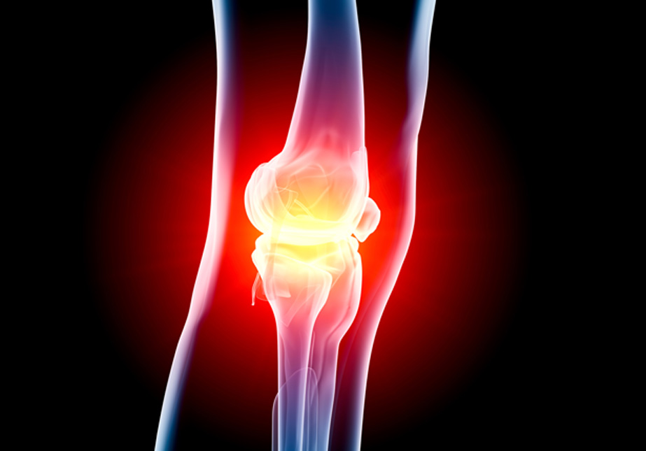
The knee naturally produces a lubricating fluid, called synovial fluid, to help the leg move smoothly and reduce friction between the different parts of the knee.
A problem with the knee joint, such as arthritis or a cartilage tear, may cause the knee to produce too much fluid and lead to a condition called Baker’s cyst. A Baker’s cyst, also referred to as a popliteal cyst, is a fluid-filled cyst that creates a bulge behind the knee.
When excess knee joint fluid is compressed by the body weight between the bones and the knee joint, it becomes trapped and separated from the joint and forms a Baker’s cyst. The cyst is named after the British surgeon who originally described the condition, William Morrant Baker.
Causes of Baker’s Cyst
The condition may be caused by:
- Damage to the knee’s cartilage (meniscus)
- Arthritis in the knee
- Osteoarthritis (also called degenerative arthritis)
- Rheumatoid arthritis
- Other knee conditions that cause joint inflammation
Risk Factors for a Baker’s Cyst
Individuals with the following conditions are at greater risk of developing a Baker’s cyst:
- Torn meniscus
- Knee arthritis (including rheumatoid arthritis and osteoarthritis)
- Knee joint injury

Symptoms of Baker’s Cyst
Signs of Baker’s cyst may include:
- Swelling behind the knee, and occasionally in the leg
- Knee pain
- Stiffness and inability to fully flex the knee
- Bruising on the knee and calf
If left untreated, the Baker’s cyst may burst and cause synovial fluid to leak into the calf region. This can cause:
- Sharp pain in the knee
- Appearance of a painless bruise under the inner ankle
- Prolonged swelling in the calf
- Redness of the calf
- Complications from related injuries, such as torn cartilage
- Feeling of water running down your calf
Diagnosing Baker’s Cyst
Patients experiencing symptoms should schedule an appointment with their primary physician, orthopedist or rheumatologist. During the initial visit, the physician will conduct a physical examination and order noninvasive imaging tests, such as an ultrasound, x-ray and MRI. The tests will help rule out other more serious conditions such as a tumor, aneurysm, deep vein thrombosis, or blood clot.
An ultrasound involves injecting a contrast dye into the knee. This is followed by imaging, called an MRI scan, or an arthrogram.
Treating Baker’s Cyst
It is possible for a Baker’s cyst to disappear on its own. But if the cyst is large and causes pain, patients may need medication, aspiration and physical therapy.
What to Expect During a Baker’s Cyst Aspiration Procedure
The patient’s affected knee is extended to make the cyst more prominent. The physician will use an ultrasound to help guide an 18- or 20-gauge needle into the correct place into the knee joint. The physician will then draw the fluid from the joint. The physician may also inject cortisone to help relieve pain and inflammation.
After the Procedure
The patient may need to undergo physical therapy to fully recover. Physical therapy may help strengthen the muscles around the knee, reduce symptoms and preserve knee function.